Scientific publications where Cell-ACDC was used
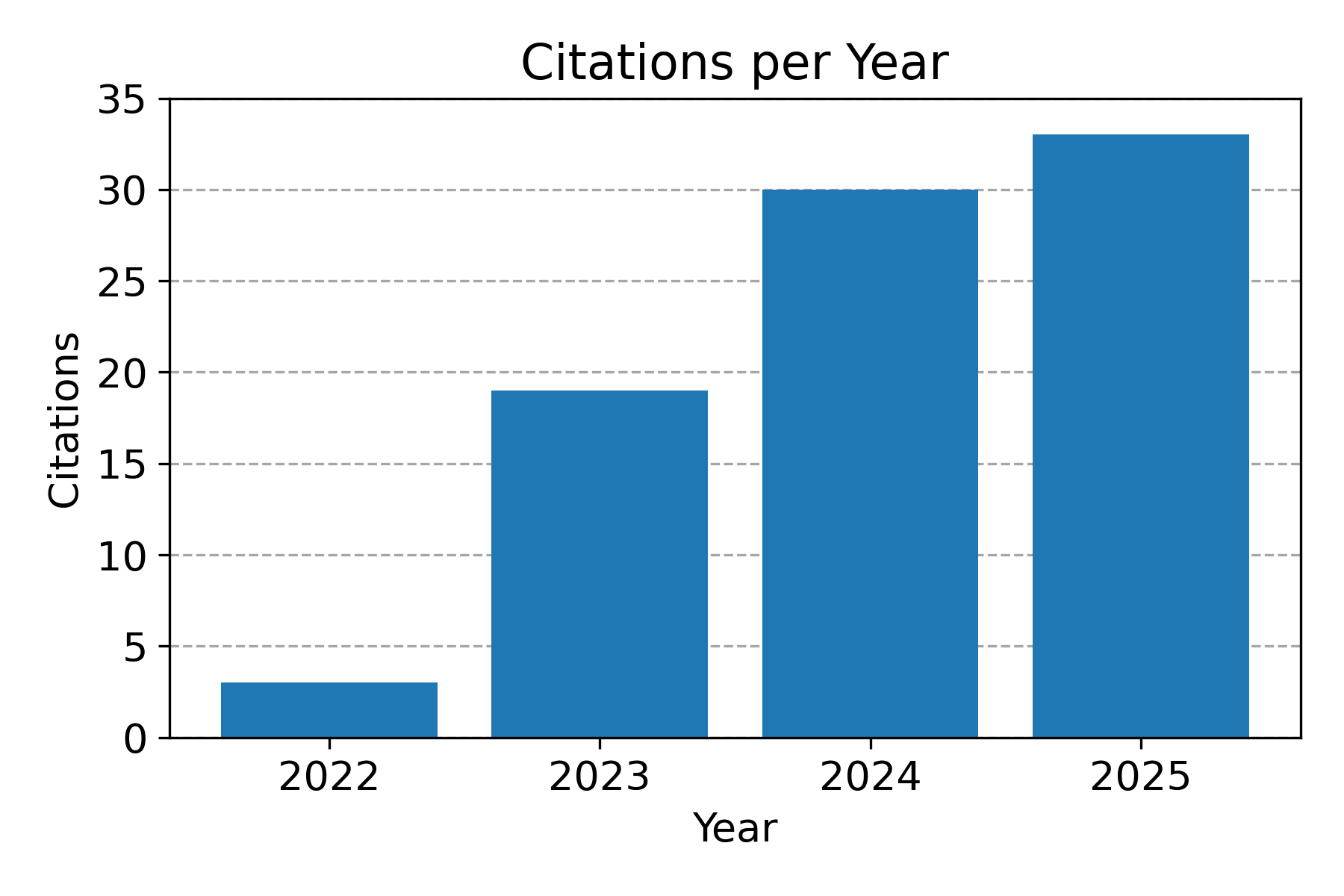
Citations per year. Data from Google Scholar.
In the following publications, authors used Cell-ACDC to analyse microscopy data and obtain biological insights:
- Moukham, H., et al. SNF1/AMPK controls its own localization by phosphorylating its activating kinase Sak1. iScience (2026) DOI: 10.1016/j.isci.2026.116357.
- Vandal, E. S., et al. The role of cell growth rate on accumulation of the mitotic cyclin Cdc13 in fission yeast. bioRxiv (2026) DOI: 10.64898/2026.05.14.724355.
- Kim, J., et al. A Fkh1/2 binding site array in the WHI5 promoter drives sub-scaling transcription. Cell Reports (2026) DOI: 10.1016/j.celrep.2026.117304.
- Benedikt, A. et al. Retrograde signalling mediates cellular adaptation to mitochondrial DNA copy number alterations. bioRxiv (2026) DOI: 10.64898/2026.04.22.720057.
- Lanz, M. C., et al. Cell enlargement drives aging-associated proteome remodeling and shortens replicative lifespan. bioRxiv (2026) DOI: 10.64898/2026.02.15.706013.
- Lambiase, A., et al. Acteoside exerts neuroprotective effects by preventing α-synuclein aggregation and oxidative stress in models of Parkinson’s disease. Neurotherapeutics (2025) DOI: 10.1016/j.neurot.2025.e00825.
- Wunder, T., et al. Conserved and Lineage-Specific Roles of KEA-Mediated Ion Homeostasis in Chlamydomonas. bioRxiv (2025) DOI: 10.64898/2025.12.03.692059.
- Gao, X., et al. Yeast growth is controlled by the proportional scaling of mRNA and ribosome concentrations. arXiv (2025) DOI: 10.48550/arXiv.2508.14997.
- Garrigós, V., et al. Tsa1-Mediated Regulation of PKA Tunes Trehalose Metabolism in Saccharomyces cerevisiae. bioRxiv (2025) DOI: 10.1101/2025.11.26.690464.
- Muñoz-Barrera, M., et al. HLH-30/TFEB is necessary for chromatin reorganization and maintenance of cell quiescence during starvation in C. elegans. bioRxiv (2025) DOI: 10.1101/2025.10.31.685810.
- Kim, J., et al. A Fkh1/2 binding site array in the WHI5 promoter drives sub-scaling transcription. bioRxiv (2025) DOI: 10.1101/2025.10.10.681508.
- Akanksha, A., et al. Microfluidic analysis of salt-stress-mediated antibiotic tolerance in Mycobacterium smegmatis. Lab on a Chip (2025) DOI: 10.1039/D5LC00713E.
- Saydee-Onwubiko, U. N., et al. Apoptosis promotes fertility in C. elegans by maintaining functional germline morphology. bioRxiv (2025) DOI: 10.1101/2025.10.22.683972.
- Proulx-Giraldeau, F. , et al. Division Asymmetry Drives Cell Size Variability in Budding Yeast. bioRxiv (2025) DOI: 10.1101/2025.10.22.683920.
- Kukhtevich, I. , et al. The origin of septin ring size control in budding yeast. The EMBO Journal (2025) DOI: 10.1038/s44318-025-00571-5.
- Conti, M. M. , et al. Dynamic phosphorylation of Hcm1 promotes fitness in chronic stress. PLOS Genetics (2025) DOI: 10.1371/journal.pgen.1011874.
- Dengler, L. , et al. When mitochondria fall apart: Unbalanced mitochondrial segregation triggers loss of mtDNA in the absence of mitochondrial fusion. bioRxiv (2025) DOI: 10.1101/2025.05.13.653688.
- Seshadri, A. , et al. Exonuclease action of replicative polymerase gamma drives damage-induced mitochondrial DNA clearance. EMBO Reports (2025) DOI: 10.1038/s44319-025-00380-1.
- Al-Refaie, N. , et al. Fasting shapes chromatin architecture through an mTOR/RNA Pol I axis. Nat. Cell Biol. 1–15 (2024) DOI: 10.1038/s41556-024-01512-w.
- Lanz, M. C. , et al. Genome dilution by cell growth drives starvation-like proteome remodeling in mammalian and yeast cells. Nat. Struct. Mol. Biol. (2024) DOI: 10.1038/s41594-024-01353-z.
- Xiao, J., Turner, J. J., Kõivomägi, M. & Skotheim, J. M. Whi5 hypo- and hyper-phosphorylation dynamics control cell-cycle entry and progression. Curr. Biol. 34, 2434-2447.e5 (2024).DOI: 10.1016/j.cub.2024.04.052.
- Vitacolonna, M. , et al. A multiparametric analysis including single-cell and subcellular feature assessment reveals differential behavior of spheroid cultures on distinct ultra-low attachment plate types. Front. Bioeng. Biotechnol. 12, (2024) DOI: 10.3389/fbioe.2024.1422235.
- Roussou, R. , et al. Real-time assessment of mitochondrial DNA heteroplasmy dynamics at the single-cell level. EMBO J. 43, 5340–5359 (2024) DOI: 10.1038/s44318-024-00183-5.
- Padovani, F. , et al. SpotMAX: a generalist framework for multi-dimensional automatic spot detection and quantification. bioRxiv (2024) DOI: 10.1101/2024.10.22.619610.
- Kukhtevich, I. , et al. The origin of septin ring size control in budding yeast. bioRxiv (2024) DOI: 10.1101/2024.07.30.605628.
- Chatzitheodoridou, D., Bureik, D., Padovani, F., Nadimpalli, K. V. & Schmoller, K. M. Decoupled transcript and protein concentrations ensure histone homeostasis in different nutrients. EMBO J. 43, 5141–5168 (2024) DOI: 10.1038/s44318-024-00227-w.
- Chadha, Y., Kukhtevich, I. V., Padovani, F., Schneider, R. & Schmoller, K. M. Single-cell imaging reveals a key role of Bck2 in budding yeast cell size adaptation to nutrient challenges. bioRxiv (2024) DOI: 10.1101/2024.10.04.616606.
- Seel, A. , et al. Regulation with cell size ensures mitochondrial DNA homeostasis during cell growth. Nat. Struct. Mol. Biol. 30, 1549–1560 (2023) DOI: 10.1038/s41594-023-01091-8.
- Piñeiro López, C., Rodrigues Neves, A. R., Čavka, I., Gros, O. J. & Köhler, S. Segmentation of C. elegans germline nuclei. MicroPubl Biol. (2023) DOI: 10.17912/MICROPUB.BIOLOGY.001062.
- Freitag, M. , et al. Single-molecule experiments reveal the elbow as an essential folding guide in SMC coiled-coil arms. Biophys. J. 121, 4702–4713 (2022) DOI: 10.1016/j.bpj.2022.10.017.
- Kukhtevich, I. V. , et al. Quantitative RNA imaging in single live cells reveals age-dependent asymmetric inheritance. Cell Rep. 41, (2022) DOI: 10.1016/j.celrep.2022.111656.
- Padovani, F., Mairhörmann, B., Falter-Braun, P., Lengefeld, J. & Schmoller, K. M. Segmentation, tracking and cell cycle analysis of live-cell imaging data with Cell-ACDC. BMC Biol. 20, 174 (2022) DOI: 10.1186/s12915-022-01372-6.
- Schuh, L. , et al. Altered expression response upon repeated gene repression in single yeast cells. PLOS Comput. Biol. 18, e1010640 (2022) DOI: 10.1371/journal.pcbi.1010640.